The Sedimentary Laboratory
Overview
The sedimentary laboratory is a centre for receiving marine sedimentary materials, carrying out non-destructive scanning techniques such as X-ray fluorescence, spectrophotometry, and magnetic susceptibility; splitting and cutting marine and other cores, and preparing samples for Mass Spectrometry, trace metal and other analyses based on the use of discrete samples. We also measure the particle sizes found in marine sedimentary deposits, as a means of investigating parameters such as changes in oceanic current speeds.
Cores
Sediments from the deep sea are retrieved in columns of sediment known as "cores", which are typically in plastic core liners. They are stored in a large refrigerated room at 4 degrees centigrade to minimise the deterioration of the cores through the action of moulds, desiccation, and other processes.
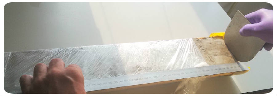
In the sedimentary laboratory we use high-frequency cutting tools to split the plastic liners, and cut and prepare the flat internal surfaces found on either side of the split core.
One side of the core is usually designated the "sampling" half. After scanning the sampling half can be sliced up into smaller - typically 1cm - subsamples for use in more destructive analyses such as isotopic and trace metal analyses.
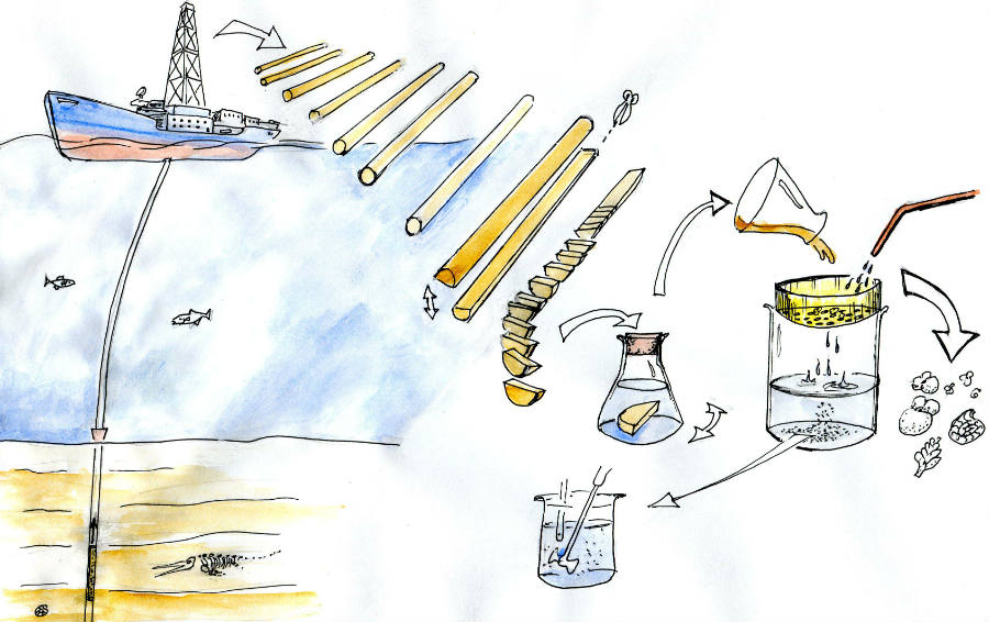
An overview of core collection and processing
X-ray fluorescence
XRF fluorescence scanning shines X-ray radiation onto the split surface of a core and returns a spectrum in the X-ray part of the light spectrum which can be resolved into changes in the relative composition of the core. Over 25 different elements are typically mapped downcore, usually at half-centimeter intervals. In this way a detailed picture of changes in core composition can be built up. Our third-generation Avaatech scanner typically takes one day to collect data on a one-and-a-half meter core.
The XRF scanner incorporates a high-quality camera which can be used to obtain detailed images of cores and other scanned materials. The images can be analysed in terms of red, green and blue wavelengths of light, and the ratios between them, and also the overall grey-level brightness of the core surface.
We use a Minolta spectrophotometer to document changes in the visible-light characteritics of the cores at a set of different light wavelengths. Changes in the abundance of heamatite and Goethite can be detected in this way, as can the extent of oxidation of the core and the presence of components such as saharan dust.
Magnetic Susceptibility
The relative contribution of magnetisable elements - "magnetic susceptibility" - can be measured using the labs Bartington MS-3 portable magnetic susceptibility meter. This again provides a non-destrucive insight into the composition of the core.
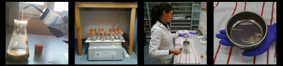
Core sampling
Cores that are ready to be split into smaller subsamples are marked along the edge of the core liner then cut with stainless-steel or plastic D-shaped cutters and placed into lebelled plastic bags. The next step is to place the sediment samples into a conical flask with approximately 100 ml of de-ionised water and then onto an orbital shaker which disaggregates the clays into a soupy slurry, which can be wet sieved.
Wet sieving
The process of wet-sieving involves pouring the samples onto a sieve - usually with a 63 micron mesh but sometimes with a 150 micron mesh, depending on the siliceous microfossil content of the sediments at the location being studied. The sieve is on top of a 1-litre pyrex flask, and as the sediments are sprayed with a fine aerosol of de-ionised water, the fine components such as clays or nannofossils are washed through the sieve into the jar beneath, where they collect as the "fine fraction" of the sample. The fine fraction is used as a resource for the study of changing proportions of (non-biological) particles in the sediment, and the geochemistry of the bulk sediment.
The fine fraction is settled in the flasks and dried out in ovens at 50 degrees centigrade for storage. Subsamples to be used for particle size analysis have their calcareous and siliceous microfossils removed by dissolution with acetic acid and sodium carbonate treatment respectively. Fine fraction can also be used for pollen analysis as it can preserve a record of terestrial changes based on the succession of particular plant species.
Coarse fraction
The coarse fraction consists of larger microfossils such as foraminifera and pteropods, and ice-rafted lithological clasts - IRD. The coarse fraction is stored in glass vials and particular species of foraminifera are picked for isotopic or trace metal analyses.
Particle size analyses
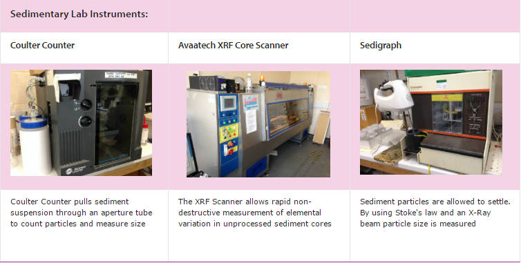
The sedimentary laboratory uses a micromeritics coulter counter mark III for particle size analyses of sortable silt, typically in the size range 4 to 63 microns. Finer constituents such as clays can be measured using the sedigraph, which uses the diffraction of X-rays in a particle settling chamber to evaluate the relative particle size composition.